Towards an Interpretable Functional Image-Based Classifier: Dimensionality Reduction of High-Density Diffuse Optical Tomography Data
Authors: Sruthi Srinivasan, Emilia Butters, Flavia Mancini, and Gemma Bale
Published: February 2024
High-density diffuse optical tomography (HD-DOT) is a wearable neuroimaging method that demonstrates high temporal and spatial resolution. While this data contains far richer information as a result, the high dimensionality and presence of complicated interconnections between data points requires the use of dimensionality reduction techniques to simplify the predictive modelling task without eliminating meaningful data features. To interrogate the possibility of designing a physiologically relevant HD-DOT feature set, cortical parcellations were applied to reconstructed images of brain activity to reduce the data dimensionality. Read more about it here.
Subject-specific information enhances spatial accuracy of high-density diffuse optical tomography
Authors: Sruthi Srinivasan, Deepshikha Acharya, Emilia Butters, Liam Collins-Jones, Flavia Mancini, and Gemma Bale
Published: February 2024
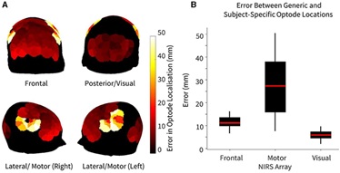
The accurate quantification of cortical activation using fNIRS data is highly dependent on the ability to correctly localize the positions of light sources and photodetectors on the scalp surface. Variations in head size and shape across participants greatly impact the precise locations of these optodes and consequently, the regions of the cortical surface being reached. Such variations can therefore influence the conclusions drawn in NIRS studies that attempt to explore specific cortical regions. In order to preserve the spatial identity of each NIRS channel, subject-specific differences in NIRS array registration must be considered. Using high-density diffuse optical tomography (HD-DOT), we have demonstrated the inter-subject variability of the same HD-DOT array applied to ten participants recorded in the resting state. We have also compared three-dimensional image reconstruction results obtained using subject-specific positioning information to those obtained using generic optode locations. Read more about it here.
A promising tool to explore functional impairment in neurodegeneration: A systematic review of near-infrared spectroscopy in dementia.
Authors: Emilia Butters, Sruthi Srinivasan, John T. O’Brien, Li Su, and Gemma Bale.
Published: September 2023
This systematic review aimed to evaluate previous studies which used near-infrared spectroscopy (NIRS) in dementia given its suitability as a diagnostic and investigative tool in this population. From 800 identified records which used NIRS in dementia and prodromal stages, 88 studies were evaluated which employed a range of tasks testing memory (29), word retrieval (24), motor (8) and visuo-spatial function (4), and which explored the resting state (32). Across these domains, dementia exhibited blunted haemodynamic responses, often localised to frontal regions of interest, and a lack of task-appropriate frontal lateralisation. Prodromal stages, such as mild cognitive impairment, revealed mixed results. Reduced cognitive performance accompanied by either diminished functional responses or hyperactivity was identified, the latter suggesting a compensatory response not present at the dementia stage. Despite clear evidence of alterations in brain oxygenation in dementia and prodromal stages, a consensus as to the nature of these changes is difficult to reach. This is likely partially due to the lack of standardisation in optical techniques and processing methods for the application of NIRS to dementia. Further studies are required exploring more naturalistic settings and a wider range of dementia subtypes. Read more about it here.
Illuminating neurodegeneration: a future perspective on near-infrared spectroscopy in dementia research
Authors: Sruthi Srinivasan, Emilia Butters, Liam Collins-Jones, Li Su, John O’Brien, and Gemma Bale.
Published: February 2023
Dementia presents a global healthcare crisis, and neuroimaging is the main method for developing effective diagnoses and treatments. Yet currently, there is a lack of sensitive, portable, and low-cost neuroimaging tools. As dementia is associated with vascular and metabolic dysfunction, near-infrared spectroscopy (NIRS) has the potential to fill this gap. Read more about it here.