Submitted by Sruthi Srinivasan on Fri, 21/04/2023 - 14:23
The Neuro-Optics lab is running a dementia study using new state-of-the art brain imaging techniques. The aim of this study is to develop new and better ways of diagnosing dementia as current methods used to detect dementia are inadequate. As a result, many cases are untreated, missed, or misdiagnosed.
To receive updates about recruitment and how to take part in this study, please enter your email into the following form: https://forms.gle/J8fXokyTEXbphbGX7.
Dementia is a global health crisis
Dementia is one of the major health issues of our time. It affects over 55 million people worldwide (World Health Organisation). As anyone who knows someone with dementia will have seen first-hand, it can have devastating effects on cognitive function, motor ability, and behaviour, and can significantly decrease the quality of life of those affected. Therapeutic interventions to lessen these effects are most promising if the dementia is diagnosed early.
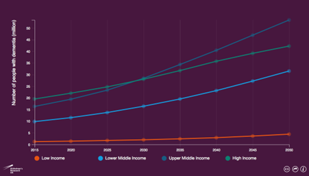
As low income countries have lower life expectancies, the prevalence of dementia is relatively low. Conversely, in upper middle income countries, as life expectancy and populations growth is higher, the prevalence of dementia is greater. From: Alzheimer's Research UK.
However, there are many problems with the way we currently diagnose dementia, meaning that it is often caught in later stages. One of the main reasons for this is the lack of adequate biomarkers for dementia. A biomarker is a molecule or characteristic that is predictive of the disease. Having objective, accessible, and effective biomarkers would mean that doctors could perform routine check-ups early-on to assess someone’s risk of developing dementia and see if intervention is necessary.
There is an urgent need to develop new biomarkers for dementia
Dementia is linked to several problems with the blood system and metabolism which are thought to occur before people even present with symptoms [1] and are strongly linked with the brain damage present. Measures of these problems may thus be good biomarkers to use to detect dementia in early stages.
Near-infrared spectroscopy (NIRS) is a neuroimaging method which uses near-infrared light to measure brain oxygenation (i.e., how well your blood system supplies the brain with oxygen) and neurometabolism (i.e., how well your brain cells use oxygen). Unlike other brain imaging techniques which you may have heard of, such as Magnetic Resonance Imaging (MRI), NIRS has several practical benefits. It is non-invasive, low-cost, and portable, and so has great potential to replace imaging techniques like MRI and better diagnose dementia.
The Optical Neuroimaging and Cognition study
To investigate how measuring the brain’s blood oxygenation and ability to use oxygen might be able to help diagnose dementia, we will scan people with different types of dementia using NIRS. This study is run by the School of Technology and the School of Clinical Medicine at the University of Cambridge, and the Cambridgeshire and Peterborough NHS Trust. This study has been developed with Professor John O’Brien and Professor Li Su, who have extensive experience performing similar clinical studies.
Who is eligible to take part?
We are looking for people with:
- A diagnosis of Alzheimer’s Disease (18 years +)
- A diagnosis of Dementia with Lewy Bodies (18 years +)
- A diagnosis of Mild Cognitive Impairment (18 years +)
- No cognitive impairment (50 years +)
- No cognitive impairment (18-50 years)
What does taking part involve?
Participating in this study will involve a single visit to the University of Cambridge, two NIRS scans, and may possibly involve an MRI scan. The first NIRS scan will use a device called LUMO (Gowerlabs Ltd) to measure brain oxygenation. The second NIRS scan will use a different device called mini-CYRIL [2] to measure brain metabolism. These scans involve wearing a special cap which contains small lights which shine onto the head. These lights are like tiny torches and are completely harmless. Both systems are completely safe. NIRS scanning is entirely non-invasive, painless, and quiet, and has very minimal, if not zero, side effects or risks to health, and is commonly used to perform brain scans on infants.
During this visit, you will be brain scanned using NIRS whilst performing various cognitive and motor tasks. You will also be asked several questions pertaining to aspects of your life, such as sleep quality and daily activities. If you have Alzheimer’s Disease or Dementia with Lewy Bodies, you may also require a separate brain (MRI) scan to look at your brain structure if you have not had one within the last 2 years.
By taking part in this study, you will be making an invaluable contribution to dementia research and helping the development of better therapeutic tools for one of the most pressing health crises affecting us today. Participation in this study is not compensated but travel expenses to and from the study site will be reimbursed. Participants will also be given access to any publications produced from the results of this study.
If you would be interested in taking part in this study or know someone who might, please follow this link (https://forms.gle/J8fXokyTEXbphbGX7) and enter your email to receive news of recruitment and information on how to get involved!
Please refer any questions to the study coordinator, Emilia Butters (emilia.butters@nhs.net).
References
- Butters et al. (2022). A systematic review of near-infrared spectroscopy in dementia. MedxRiv. Doi: 10.1101/2022.11.23.22282361.
- Bale et al. (2014). A new broadband near-infrared spectroscopy system for in-vivo measurements of cerebral cytochrome-c-oxidase changes in neonatal brain injury. Biomed Opt Express. 5(1): 3450-66. Doi: 10.1364/BOE.5.003450.